The University Hospital Bonn has introduced a groundbreaking mixed-reality system for thoracic surgery, integrating holographic overlays and real-time 3D imaging to enhance surgical planning for chest wall procedures. This advanced approach, described in a study titled "A System for Mixed-Reality Holographic Overlays of Real-Time Rendered 3D-Reconstructed Imaging Using a Video Pass-through Head-Mounted Display—A Pathway to Future Navigation in Chest Wall Surgery," published in J. Clin. Med. 2024, aims to improve surgical outcomes and patient care by providing detailed preoperative visualization of a patient's anatomy.
https://doi.org/10.3390/jcm13072080
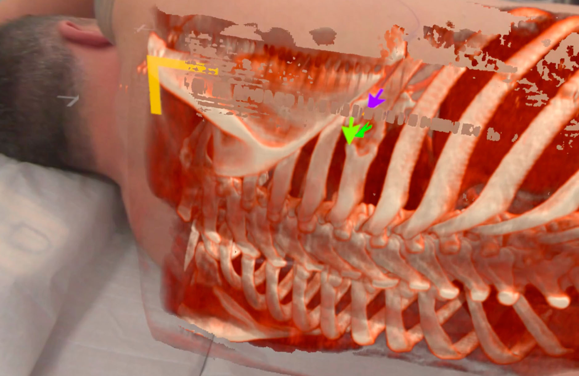
Innovative Surgical Planning
The core of this innovation involves high-resolution, three-dimensional imaging to project a holographic representation of the patient's anatomy onto their body, enabling surgeons to visualize and interact with anatomical structures in real time. This method differs significantly from traditional preoperative planning, which often relies on 2D imaging or static 3D models. The system uses a video pass-through head-mounted display connected to a high-performance workstation, allowing real-time rendering and manipulation of images.*
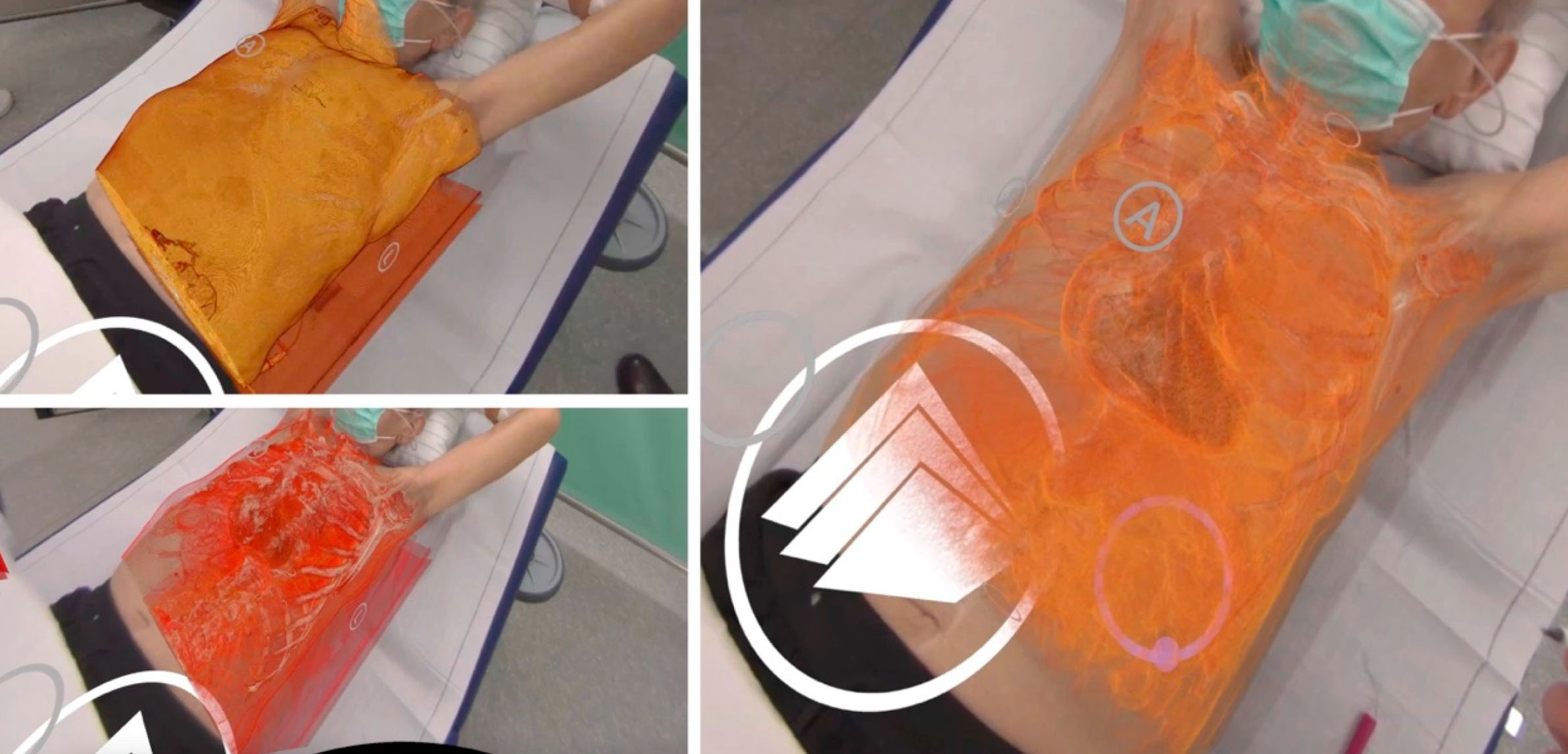
Demonstrated Efficacy
The technology's efficacy was demonstrated through three oncological cases, highlighting its potential to offer invaluable insights into the spatial relationships between tumors and critical anatomical structures, facilitating informed surgical planning. Looking ahead, this technology could be integrated into actual surgical procedures, providing real-time guidance and navigation, thereby reducing reliance on intraoperative imaging and minimizing radiation exposure.
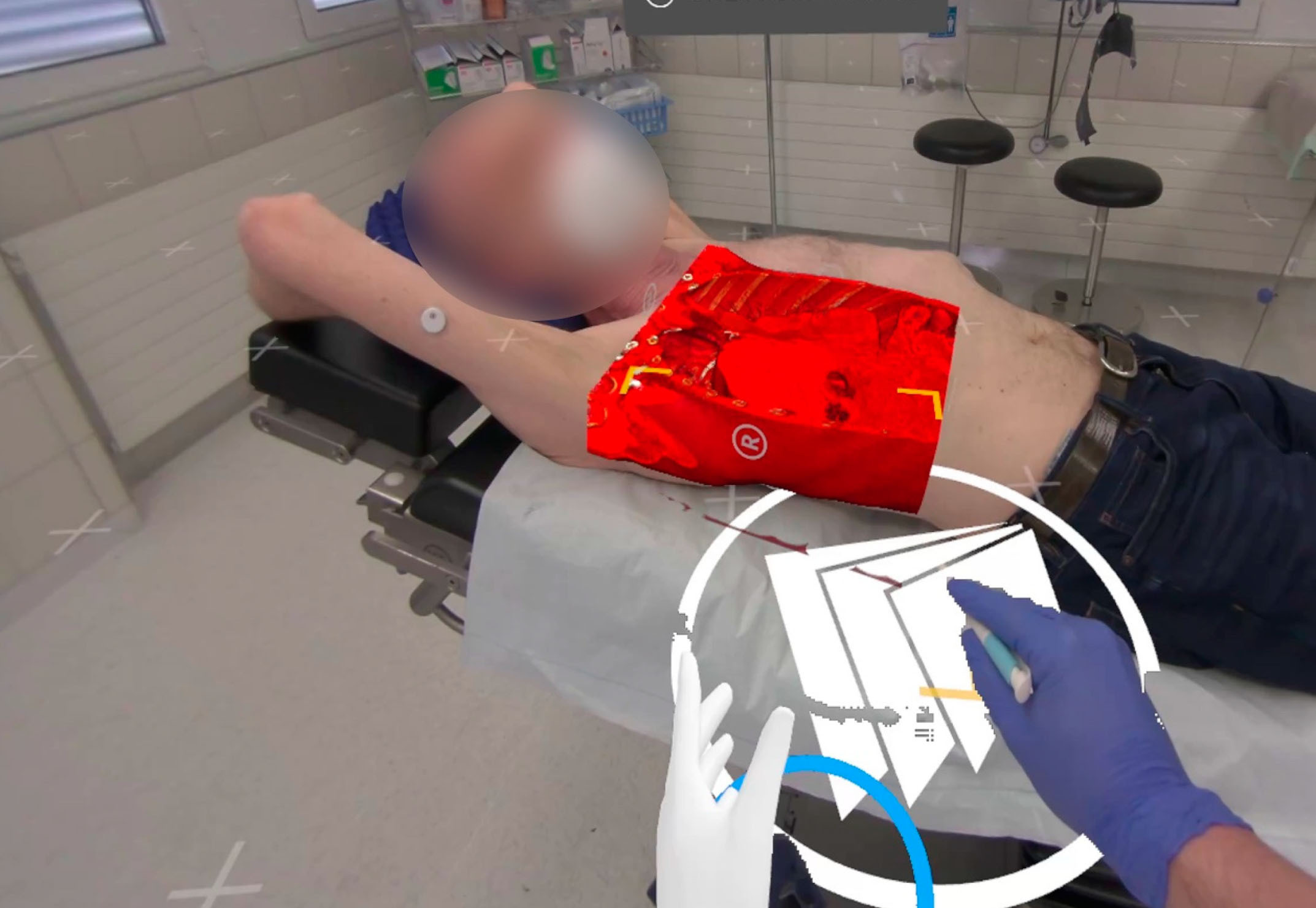
Overcoming Challenges and Future Outlook
Despite the promising advancements, challenges remain, such as the system's tethered connection to a workstation and regulatory hurdles, particularly for intraoperative use. However, with ongoing developments in mixed-reality hardware and cloud computing, these obstacles are expected to decrease, making such sophisticated systems more accessible for surgical applications. This study emphasizes the significant potential of mixed-reality and real-time 3D imaging in revolutionizing surgical planning and execution, urging further research to fully explore its capabilities, accuracy, and impact on clinical outcomes in thoracic surgery and potentially other fields.
*Important Notice Regarding Functionalities for Surgical Planning and Professional Clinical Usage
The specialized features tailored for surgical planning and pre-operative professional applications are exclusive to Medical Imaging XR PRO. At the moment, Medicalholodeck's applications are only available for educational purposes.